


Microstructure de la neige
La thématique Microstructure de la Neige concerne l’étude de la structure de la neige à l’échelle de ses grains de glace. Elle s’appuie sur des outils expérimentaux et numériques tels que la microtomographie, l’analyse d’images et la simulation numérique.
Personnel impliqué actuellement :
– Responsable de la thématique : Frédéric Flin
– Membres permanents : Neige Calonne, Pascal Hagenmuller
Anciens membres :
– Membres permanents : J.-B. Brzoska, C. Coléou, Anne Dufour, B. Lesaffre, P. Lapalus, L. Pézard
– Etudiants et Post-docs : P. Bertrand, S. Borel, L. Bouvet, S. Cabanes, N. Calonne, R. Caneill, P.-J. Font, L. Gillibert, R. Granger, I. Haffar, A. Hasan, P. Latil, C. Mehu, I. Peinke, A. Philip, R. A. Pieritz, A. Regenscheit, B. Voisin, X. Wang, A. Wautier, A. Zennoune
Principales collaborations :
– Tomographie : 3S-R Lab, ESRF, SOLEIL
– Neige et glace : ILTS, IPAG, INRAE / Etna, IGE
– Analyse d’images : HIT, LAMA, LIRIS
– Modélisation : 3S-R Lab, ICJ, LAMA, IGE, LJK
– Applications : 3S-R Lab, Airbus, IFTS, INRAE / FRISE, LaMCoS
Etudes à microéchelle
During a snowfall, the snow crystals accumulate on the ground and gradually form a complex porous medium constituted of air, water vapor, ice and sometimes liquid water. This ground-lying snow transforms with time, depending on the physical parameters of the environment. This process, called metamorphism, can be divided into three main types of metamorphisms : the wet snow metamorphism, the isothermal metamorphism, and the temperature gradient (TG) metamorphism. In polar or mountainous conditions, these processes are followed by compaction of snow into ice under the load of the upper snow layers.
Although the effects of theses different transformations are roughly well-known, the physical mechanisms that lead to these transformations are not yet perfectly well understood. A better comprehension of these mechanisms would help understanding how the snow and ice change their microstructure depending on the applied physical conditions. For instance, this offers interesting outcomes to forecast snow, firn and ice thermal, mechanical and chemical properties.
This research has potential major impacts in different research fields such as :
– avalanche risk forecasting ;
– artificial snow management ;
– environmental and atmospheric chemistry ;
– past climates and interpretation of ice cores ;
– numerical modeling of the cryosphere ;
– snow road viability.
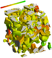
Etudes tridimensionnelles
In most of common microstructural studies, materials are generally observed with the help of 2D projections (optical microscopy, SEM imaging, ...). These kind of observations are generally easy and fast, and allow the investigation of a large amount of samples. However, these images are often difficult or impossible to use quantitatively, because important information is missing : materials volumes generally have a complex three-dimensional structure, while the investigation process is only bi-dimensional.
Hopefully, new techniques such as confocal imaging or tomography are becoming easily available and allow to obtain precise three-dimensional information from the inside of the investigated samples, without damaging the sample. By using image analysis algorithms, it is then possible to extract a lot of information from these 3D original images. For instance, such images can :
– provide accurate and complete quantitative data measurements that are not biased by projection effects ;
– give access to typical 3D key parameters such as local curvature, Euler number and other connectivity-related parameters that should be necessarily considered in 3D ;
– open new fields to the three-dimensional simulation of physical processes because numerical models can be applied directly on the numerical 3D image of real samples.